
Computed tomography (CT) of the heart
Everything you need to know about the examination.
In a cardiac CT scan, the coronary arteries themselves are examined for narrowing (coronary stenosis) and the extent of coronary artery calcification (degree of coronary sclerosis).
This gentle examination technique of the coronary arteries has recently been recommended for certain patients if narrowing of the coronary arteries is suspected. This means that a cardiac catheter examination via the groin or arm (so-called invasive coronary angiography) can often be avoided.
Frequently asked questions
How can I prepare for the examination?
What you can do yourself:
You do not need to fast before a computed tomography (CT) scan of the heart. You should also take your usual medication (tablets) as normal. The scan requires the intravenous administration of an iodine-containing contrast agent. We therefore require patients aged 60 and over (≥ 60 years) to provide a current creatinine and TSH level on the day of the examination, which should not be more than four weeks old. These two values provide information about your kidney and thyroid function and can, for example, be determined in advance by your GP or the referring doctor via a blood test. If you have ever had an allergic reaction to a CT contrast agent in the past or noticed any intolerance (e.g. skin rash, breathing difficulties, circulatory reactions), please be sure to inform us of this at the time of booking. This information is very important to us so that we can plan your examination safely and take preventive measures if necessary.
To obtain the clearest possible CT images of the heart, a relatively low heart rate (approx. 55–65 beats per minute) is important. Please therefore avoid consuming any food or drink containing caffeine or theine for 12 hours prior to the examination, as these can increase your heart rate.
How your GP/cardiologist can best prepare for the examination:
The attached checklist serves as a guide for medical colleagues and patients to rule out contraindications for the necessary administration of contrast agents, beta blockers and nitro spray. This is important to ensure that the cardiac CT examination runs as quickly, smoothly and safely as possible. If necessary, the listed medications should only be discontinued after consultation with the attending physician.
Please bring the completed checklist with you to your examination appointment.
How does the examination work?
In order to obtain the sharpest possible images of the heart, it may be necessary to lower your heart rate (pulse) by administering additional medication (ß-blockers). To further improve the assessability of the coronary arteries, you will also be given two sprays of a medication (nitro spray) under your tongue. This causes the coronary arteries to dilate, thereby increasing the accuracy of the detection of relevant narrowings. Both medications are safe if there are no contraindications and are generally very well tolerated by patients. The radiologist on site will discuss this with you in detail and will of course be available to answer any questions you may have.
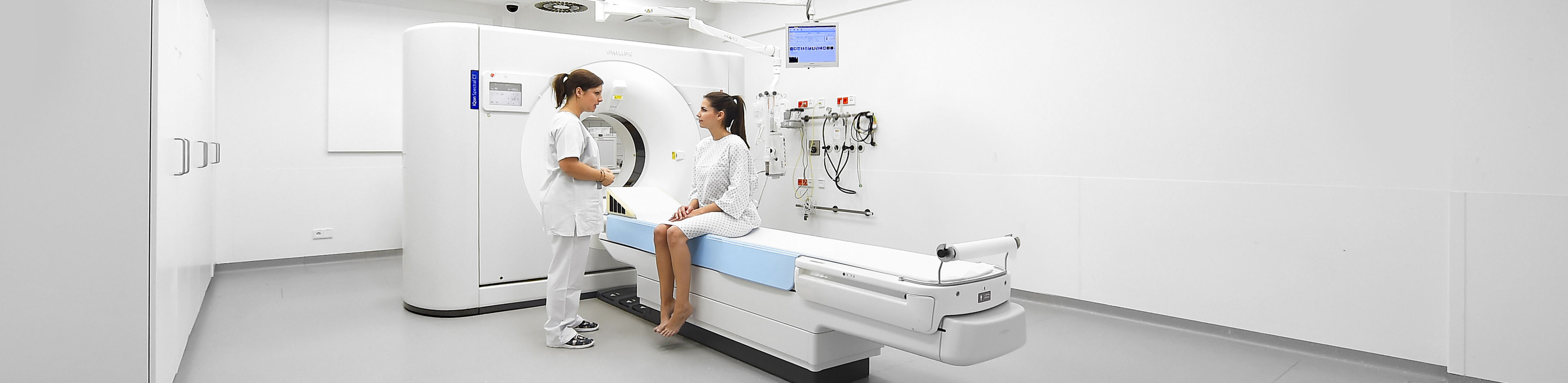
Preparations for the examination take about 20-25 minutes, and the examination itself takes only a few seconds. During the examination, you will be positioned on the examination table with your arms raised above your head so that your chest area can be examined. You will then be moved into the computer tomograph (CT) with your feet first on the table. The modern examination equipment commonly used today has a very wide opening (more like a large arch), so claustrophobia is not usually a problem.
In order to administer the contrast agent and beta blocker, we will need to insert an intravenous line into a superficial vein in your arm before the examination, which is similar to having blood taken. The contrast agent is administered via an automated pump during the examination. You may feel a little warm, which is completely normal.
After the cardiac CT scan is complete, you can resume your normal daily activities. The CT scan is then evaluated and assessed by radiologists who are specially trained and experienced in cardiac imaging and certified by the German Radiological Society in the field of cardiac CT. To do this, the entire heart and the individual coronary arteries are assessed and examined for the presence of narrowings or other abnormalities. In addition to conventional 2D data, a 3D model of the heart is also created for better visualisation. We also use advanced reconstruction algorithms and virtual monoenergetic images of the spectral data sets to reduce any beat or blooming artefacts and increase vascular contrast.
If you would like to receive a CD or online access to the images from your examination, we will be happy to provide you with these after the examination. If you would like a final consultation with one of our radiologists to discuss the findings, this is of course possible. Please allow for some waiting time, as the images must first be evaluated. The final findings are usually prepared on the same day and sent to your treating physician.
After the examination
After the cardiac CT scan, you can go about your daily routine as normal. The CT scan is then evaluated and assessed by radiologists who are specially trained and experienced in cardiac imaging and certified by the German Radiological Society in the field of cardiac CT. To do this, the entire heart and the individual coronary arteries are assessed and examined for narrowings or other abnormalities. In addition to conventional 2D data, a 3D model of the heart is also created for better visualisation. We also use advanced reconstruction algorithms and virtual monoenergetic images of the spectral data sets to eliminate any beat or blooming artefacts and increase vascular contrast.
If you would like to receive a CD or online access with the images from your examination, we will be happy to provide you with these after the examination. If you would like a final consultation with one of our radiologists to discuss the findings, this is of course possible. Please allow for some waiting time for this, as the images must first be evaluated. The final findings are usually prepared on the same day and sent to your treating physician.
Further information
- Information on our areas of focus
- Technical details about the equipment of our institute
- Find out more about computed tomography research at our institute here