
Frequently asked questions
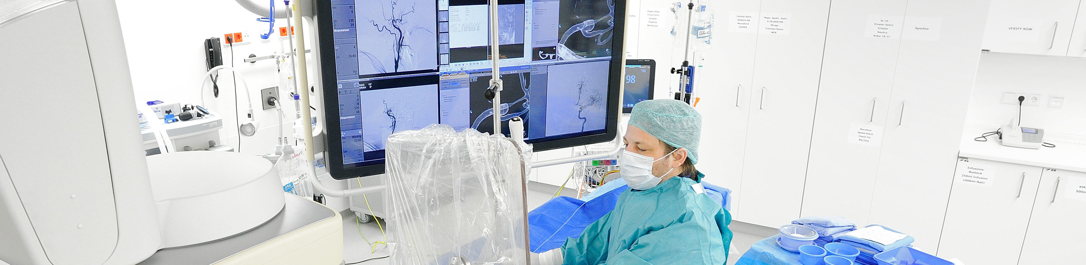
Angiographic examination is the invasive (i.e. associated with vascular puncture) visualization of vessels by direct administration of contrast medium. Angiography is usually performed as digital subtraction angiography (DSA). This also offers the possibility of additional treatment of angiographic findings (e.g. vascular constrictions).
Catheter-supported angiography at our institute is generally associated with a one-day inpatient stay. Please make an appointment via our central registration desk. On the day before the procedure, please first register at our central registration desk on the first floor of the DTZ. There you will receive a "routing slip", which you should use to be admitted as a pre-inpatient at the central patient admission desk in the entrance area of the ward block. To do this, you will need a "referral for an inpatient stay" from your referring doctor and your health card. After successful pre-inpatient admission, you will then come back to our registration desk in the DTZ, which will forward you to the angiography department. There you will then have an informative discussion with one of our radiologists. They will inform you about the benefits and risks of the procedure. You will receive a dedicated information sheet from us to prepare for the consultation. We ask you to read this carefully and complete it truthfully. You can then ask your questions during the subsequent consultation.
Please bring a current creatinine and TSH value as well as a coagulation laboratory (haemoglobin, haematocrit, platelet count, Quick value, PTT) with you to the consultation, which you will have determined in advance (about one week before the procedure) with a blood test from your family doctor or the referring doctor. These values provide information about your kidney and thyroid function as well as your coagulation status. You will be asked to present these values during your consultation with one of our radiologists. If any of the values are abnormal (i.e. not normal), please inform us in advance so that we can make the necessary decisions and plan the examination.
If you are taking blood-thinning medication, please let us know when you make your appointment. If you have a CD with externally produced images of the vessels to be treated, please also bring this with you to the consultation.
A light diet is permitted on the evening before and on the day of the procedure. You should also take medication, e.g. for cardiac arrhythmia or high blood pressure, on the day of the examination. If you are unsure about taking medication, please ask your doctor or radiologist during the consultation.
On the day of the procedure, you will be admitted to ward 4.1 in the Heart Center (vascular surgery). We will then call you from there to perform the procedure. During the procedure, after thorough disinfection and under local anesthesia, we will insert a special puncture needle (diameter approx. 1 mm) into an easily accessible artery or vein. A thin, very flexible wire is advanced through the puncture needle into the inside of the blood vessel. A catheter (i.e. a flexible plastic tube) is then inserted over this guide wire. In order to make the blood vessels visible and assessable, contrast medium is injected into the affected vessels via the catheter. During the injection of contrast medium, a feeling of warmth usually occurs in the examined body region; this is normal and only lasts a few seconds. The contrast medium is generally very well tolerated; only in rare cases can there be side effects, about which the doctor will inform you in detail. During the injection of the contrast medium, X-ray images of the affected examination region are taken in quick succession. If your kidney function is impaired, we also have the option of applying CO2 for vascular contrasting. This allows the amount of iodine-containing contrast medium to be applied to be reduced, in some cases significantly, and the risk of further deterioration in kidney function as a result of the contrast medium administration can be reduced.
The examination is usually completed after 15 to 30 minutes. Complicated anatomical conditions or the implementation of a therapeutic measure can lead to an extension of the examination time. After removing the catheter material, the puncture site is compressed for around ten minutes and then a pressure bandage is applied. Alternatively, there are also special sealing systems that immediately seal the puncture site. However, it is not always possible to use them, which is why the doctor must decide on a case-by-case basis whether a sealing system or a classic pressure bandage should be used to stop the bleeding.
After the examination, please adhere to the recommendations regarding bed rest (which will be determined by the attending radiologist) and do not remove the pressure bandage prematurely. You will usually be discharged the day after the procedure (after complications have been ruled out using ultrasound). A written report will be sent to the referring practice. Avoid heavy physical exertion for about two days. The risk of secondary bleeding with possible bruising can thus be largely excluded.
Inform your doctor immediately if you experience nausea, diarrhea, malaise, skin rash, increasing pain, fever (over 38 °C) or chills, blistering and/or a feeling of coldness or numbness in the affected arm/leg after the examination, paralysis, impaired vision, speech or hearing and other symptoms, even if these symptoms only occur a few days after the procedure! If she/he cannot be reached, please call an emergency doctor if you have serious symptoms.
Further information
- Information on our areas of focus
- Technical details on the equipment at our institute
- Research in interventional radiology