

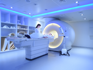
Magnetic resonance imaging, also known as magnetic resonance imaging, allows the structure and function of internal organs and soft tissue to be visualized. It is based on very strong magnetic fields and alternating magnetic fields in the radio frequency range. Even minor changes in the body, such as small areas of inflammation, can be detected in this way. In contrast to computer tomography, MRI does not require X-rays and is a very low-risk examination procedure.
Thanks to high resolution and high tissue contrast, anatomical structures can be visualized much better than with most other imaging techniques. In particular, different soft tissue structures can be better differentiated. To date, no damage has been reported due to the high magnetic flux density and electromagnetic high-frequency fields that occur during MRI. At our institute, we work with MRIs with field strengths of 1.5 and 3.0 Tesla. Find out more about magnetic resonance imaging.
Further information
- Learn more about MRI research at our institute
- Clinical MRI procedures offered at our institute