
MRI-guided vacuum biopsy of the breast
Everything you need to know about the procedure.
This minimally invasive biopsy technique is performed if an abnormality, for example a suspected breast carcinoma (breast cancer), can only be detected by MR mammography, but not by X-ray mammography or ultrasound. The indication is made by the radiologist in close cooperation with the treating gynecologist. The procedure is performed on an outpatient basis.
Frequently asked questions
MRI-guided vacuum biopsy of the breast (mamma) is a procedure in which a tissue sample is taken from the breast. The tissue sample (biopsy) is taken with a special biopsy needle under local anesthesia. The procedure is performed after an MRI scan so that the medical staff know exactly where the tissue to be removed is located inside the breast and where to insert the needle. The procedure is used for the diagnosis and early detection of breast cancer (breast carcinoma).
You do not need to be fasting for the procedure. Please do not take any blood-thinning medication five days before the procedure (this also includes the commercially available "painkillers" Aspirin©). If you are taking Heparin or Marcumar©, please let us know in advance so that we can plan the examination together with the radiologist carrying out the procedure.
Before the procedure, you will be informed by a radiologist about the benefits and risks of the procedure. You will receive a dedicated information sheet from us in preparation for the consultation. We ask you to read this carefully and complete it truthfully. You can then ask your questions during the subsequent consultation.
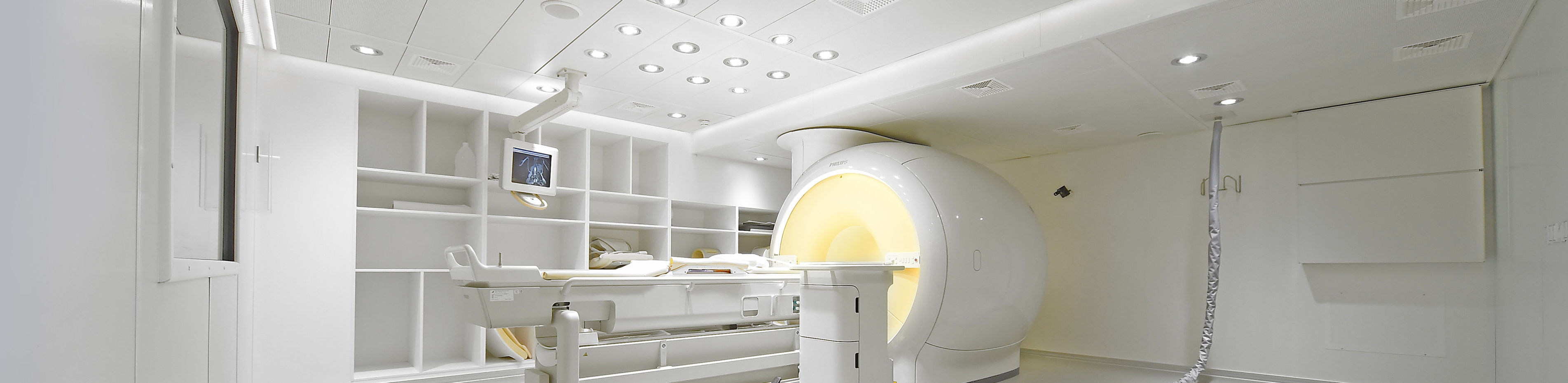
The intervention takes place in the prone position in one of our MR tomographs. The breast to be examined is slightly compressed in a special biopsy coil during the procedure. First, a normal MR mammogram is performed with intravenous administration of contrast medium, as you are already familiar with from the previous examination. Only in this way is it possible to visualize the suspicious findings. For this purpose, we will place a venous access in your arm (crook of the elbow or back of the hand) before the procedure. We only use the latest generation of contrast agents, which are very well tolerated; only in rare cases can there be side effects, about which your doctor will inform you in detail. The tissue removal (biopsy) is then planned and carried out with software support based on the examination performed.
The biopsy itself is then carried out outside the "tube". First, the skin is thoroughly disinfected and the puncture site is anesthetized locally. The biopsy needle is then inserted into the breast at the exact location of the suspicious lesion and targeted samples are taken. A small metal clip is then inserted to mark the biopsy site. The total duration of the procedure, including preparation and follow-up, is around 45 to 60 minutes. You can then go home after a short follow-up period. Please take it easy for 24 hours after the procedure to minimize the risk of post-operative bleeding.
Further information
- Information on our areas of focus
- Technical details about the equipment at our institute
- Research in interventional radiology