
Periradicular therapy
What you should know about the therapy method.
If you have been referred to us for periradicular therapy (PRT), you can find out about the procedure in advance here. Links for referring physicians, consultation requests, etc. can be found at the bottom of the page.
Periradicular therapy (PRT) describes the image-guided, percutaneous application (application through the skin) of medication directly to a nerve root shortly after it emerges from the spinal canal.
A distinction is made between so-called "therapeutic PRT" to relieve pain that occurs as a result of compression or irritation of a nerve root and "diagnostic PRT", which aims to identify the level at which surgical relief is most likely to lead to pain relief if the symptoms are unclear, for example as a result of disc protrusions at several levels.
During PRT, a local anaesthetic is administered via a thin cannula together with an anti-inflammatory medication if required. Computer tomography (CT) is used to ensure precise application directly to the irritated nerves. With the right indication, patients can sometimes experience an improvement in their symptoms after the first treatment. However, it is often necessary to repeat the treatment at intervals of several weeks before the patient is finally free of symptoms. In the case of neurological deficits, however, PRT is by no means a substitute for surgical relief of the damaged nerve root.
Approximately one day before the procedure, you will be informed by a radiologist about the benefits and risks. In preparation for the consultation, you will receive a detailed information sheet from us. We ask you to read this carefully and complete it truthfully.
You can then ask your questions during the subsequent consultation. It is not necessary for you to be sober for a PRT.
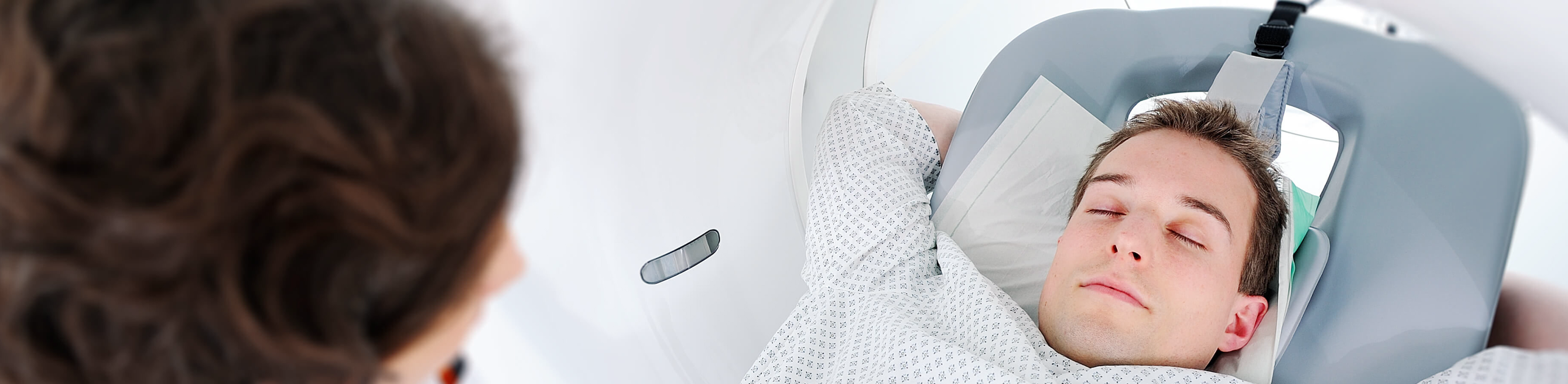
You are wheeled into the CT scanner. The modern examination devices in use today have a very wide opening (more like a large arch), so that there is usually no reason for claustrophobia. Several images are taken, which are used by the radiologist to plan the procedure. This is followed by intensive skin disinfection and marking of the puncture site. The thin needle is then brought up to the nerve root to be treated under repeated image control. After a final image-guided position check, the local anesthetic is then applied. The total duration of the procedure, including preparation and follow-up, is around 20-30 minutes.
Further information
- Find out more about neuroradiology research at our institute here
- Are you a referring physician or do you need a second opinion? Find out more under options for referring physicians
- Information on consultation requests from practices or external hospitals
- Read more about our equipment here
- Interventional radiology as a focus at the institute
- Research in interventional radiology