
X-ray stereotactic vacuum biopsy of the breast (mamma)
Everything you need to know about the procedure.
The minimally invasive X-ray stereotactic biopsy technique is used to clarify abnormal changes in the mammogram, especially microcalcifications, and is used for the diagnosis and early detection of breast cancer (breast carcinoma). The indication is given by the radiologist in close cooperation with the treating gynecologist. The procedure is performed on an outpatient basis.
Frequently asked questions
X-ray stereotactic vacuum biopsy of the breast (mamma) is a procedure in which a tissue sample is taken from the breast. The tissue sample (biopsy) is taken with a special biopsy needle under local anesthesia. The procedure is carried out after an X-ray mammogram so that the medical staff know exactly where the tissue to be removed is located inside the breast and where to insert the needle. The procedure is used for the diagnosis and early detection of breast cancer (breast carcinoma).
You do not need to be fasting for the procedure. Please do not take any blood-thinning medication five days before the procedure (this also includes the commercially available "painkillers" Aspirin©). If you are taking Heparin or Marcumar©, please let us know in advance so that we can plan the examination together with the radiologist carrying out the procedure.
Before the procedure, you will be informed by a radiologist about the benefits and risks of the procedure. You will receive a dedicated information sheet from us in preparation for the consultation. We ask you to read this carefully and complete it truthfully. You can then ask your questions during the subsequent consultation.
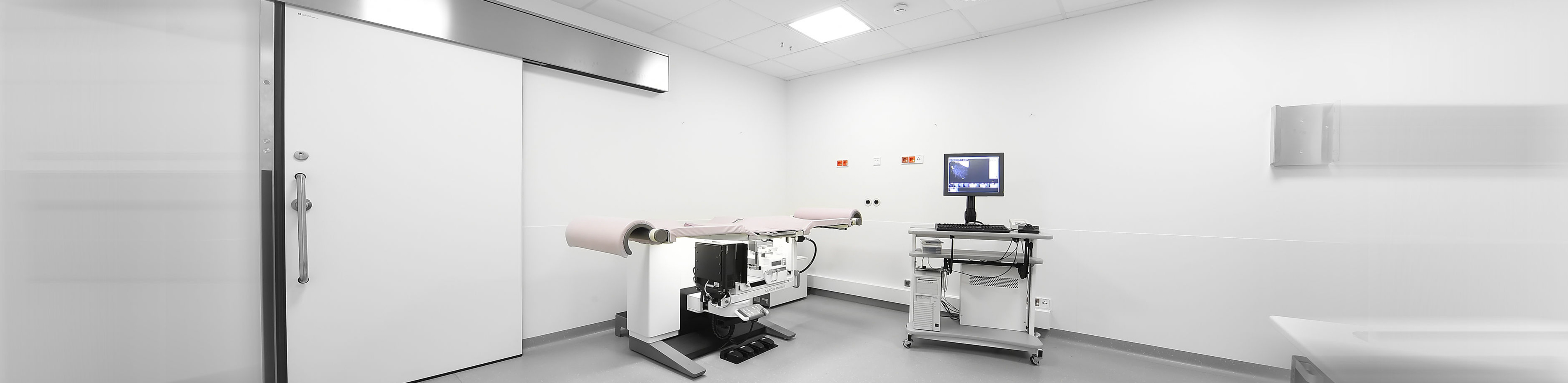
The intervention takes place in the prone position on the biopsy table. The breast to be examined is slightly compressed during the procedure. First, a few images of a small part of the breast are taken using a mammography device integrated into the table. The biopsy is then planned and carried out on the basis of these images. First, the skin is thoroughly disinfected and the puncture site is anesthetized locally. The biopsy needle is then inserted into the breast at the exact location where the conspicuous changes are located and samples are taken. It is often necessary to insert a small metal clip to mark the biopsy site, especially in the case of small findings. The total duration of the procedure, including preparation and follow-up, is around 45 to 60 minutes. You can then go home after a short follow-up period. Please take it easy for 24 hours after the procedure to minimize the risk of post-operative bleeding.
Further information
- Information on our areas of focus
- Technical details about the equipment at our institute
- Research in interventional radiology