Contact
Appointments Office
Tuesdays and Thursdays from 09:00 - 12:00
Phone +49 221 478-82035
E-mail neuroradiologie@uk-koeln.de
Building 18a Site plan
Frequently asked questions
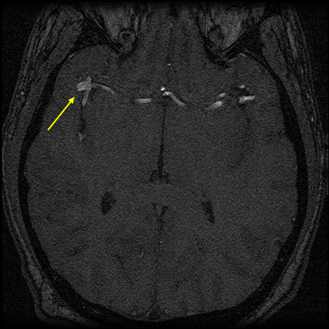
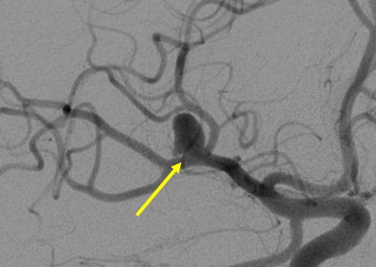
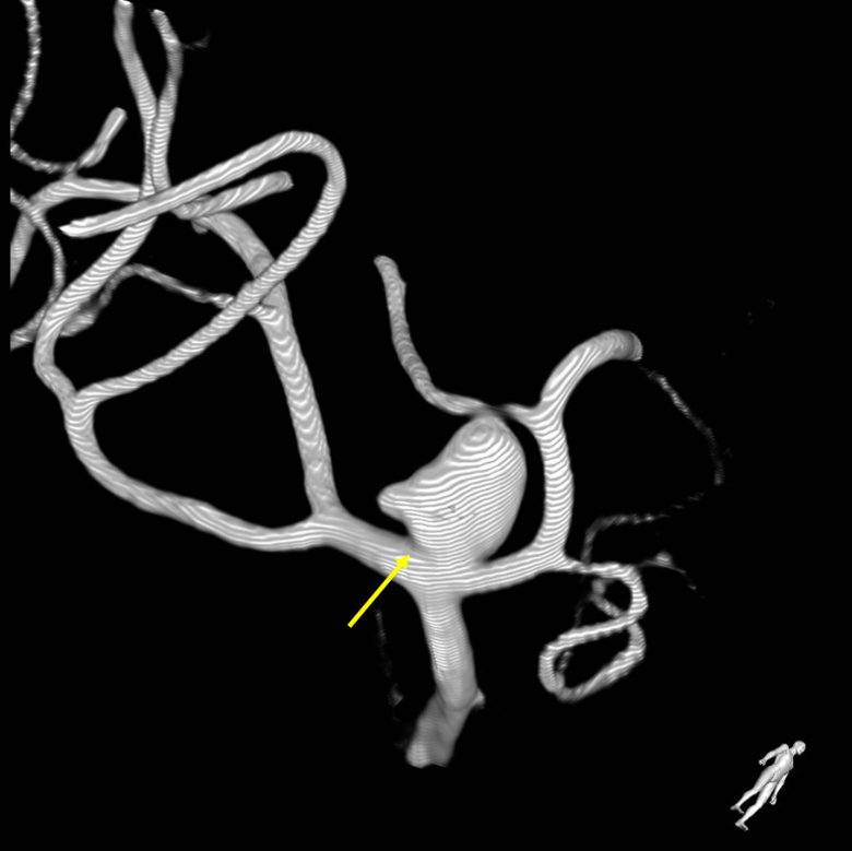
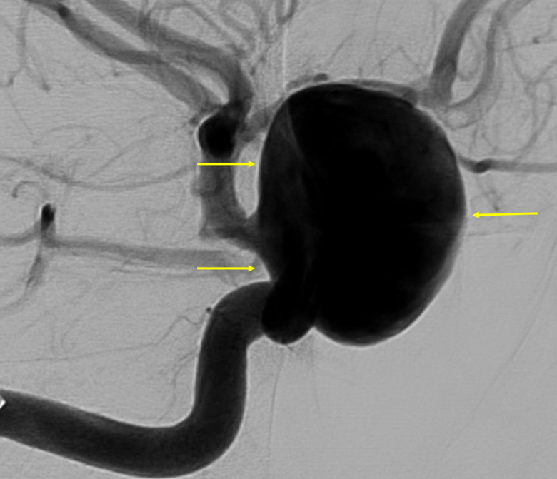
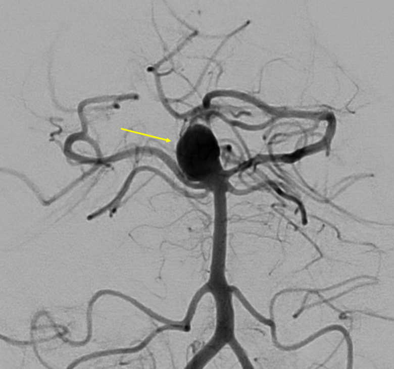
The blood vessels that supply the brain can develop bulges - so-called aneurysms. As a rule, you are not born with an aneurysm, but with a predisposition to a weak spot in the vessel wall. Over the years, a weak spot can develop into an aneurysm. This happens particularly frequently in places where the vessel branches out, as the blood flow is divided there and additional strain is placed on the vessel wall. This process is also facilitated by factors such as high blood pressure or nicotine consumption.
Aneurysms often occur spontaneously; however, there are also familial forms. In patients with more than two aneurysms, first-degree relatives should be examined by MRI. The same applies if an aneurysm has been detected in two or more first-degree relatives or in identical twins where one twin has an aneurysm.
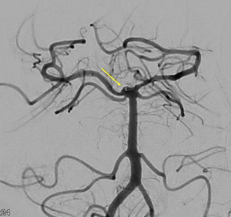
Not every aneurysm needs to be treated. Whether treatment is necessary depends on many factors. Not only the size and location of the aneurysm play a role, but also the external shape. In order to provide comprehensive advice, an additional examination using the most precise technical procedure, catheter angiography, is often necessary - this is still the gold standard in aneurysm diagnostics.
The probability depends very much on the location and shape of the aneurysm. Aneurysms in the arteries that supply the cerebellum and brain stem bleed at smaller sizes than aneurysms in the internal carotid artery, for example. The same applies to aneurysms in the anterior cerebral artery. In general, a bleeding risk of 2 to 4% per year can be assumed. Age therefore plays an important role in the individual consultation on possible treatment.
If bleeding occurs from an aneurysm, the leak usually closes quickly on its own due to a blood clot. If this does not happen, the patient is usually not admitted to hospital. The more frequent case, however, is the leakage of blood with subsequent closure of the aneurysm by itself. The blood enters the so-called subarachanoid space (space filled with cerebrospinal fluid (CSF) between the meninges and the surface of the brain); the bleeding is therefore called subarachnoid hemorrhage. The patient's symptoms range from severe headaches ("like never before") with dizziness and nausea to immediate coma. The risk of further bleeding is very high in the first few days after a subarachnoid hemorrhage. As this secondary bleeding is often not survived, treatment of the aneurysm is necessary. The patient first undergoes diagnostic angiography. The case is then discussed with colleagues from the Department of Neurosurgery and, if necessary, Neurology and the best individual form of treatment is determined.
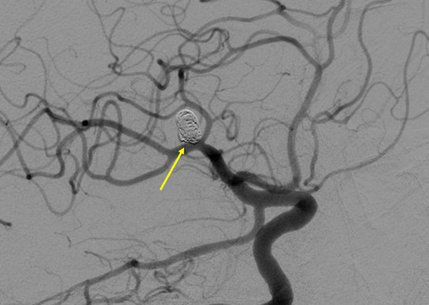
During aneurysm treatment with coils (platinum spirals), the aneurysm is treated in a minimally invasive manner from "inside" through the blood vessels. The patient is under general anesthesia, not because pain could occur, but to prevent any movement of the patient during the treatment. Access is gained through the leg artery just below the groin. A thin tube (catheter) is used to probe the aneurysm. Several platinum coils are inserted through this into the aneurysm. Ideally, this leads to 40 to 50% filling of the aneurysm with coil material and thus to a slowing of the blood flow in the aneurysm. This forms a stable complex of platinum coils and organized blood. The aneurysm is then eliminated from the blood circulation.
Coils are constantly being further developed and are available from us in over 200 different shapes and materials; always compiled according to the latest scientific findings. Ideal for "pure" coiling is when the aneurysm sits narrowly on the blood vessel, i.e. is teardrop-shaped in the broadest sense. The coils can then be firmly anchored in the aneurysm. If the contact surface of the aneurysm is wider, it may make sense to insert a stent to artificially narrow the contact surface and stabilize the coils in the aneurysm. Stents are also available in many different shapes and types.
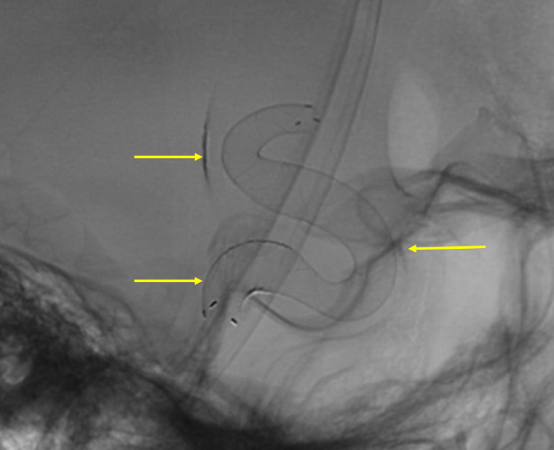
For some aneurysms, treatment with a so-called flow diverter ("flow divider") makes sense. This is a stent with a very narrow mesh density. This means that the blood flow hardly reaches the aneurysm. The altered flow and pressure conditions cause the blood in the aneurysm to clot, which then shrinks over the course of months.
Another treatment method that can be useful in some cases is the use of flow dividers in the aneurysm (so-called WEB devices). In this procedure, instead of several coils being inserted into the aneurysm, a single implant is inserted, which also diverts the blood flow out of the aneurysm.