
Heart MRI (Cardio MRI)
Magnetic resonance imaging (MRI) is the best and most accurate method for examining the heart for many issues and does not require the use of ionizing radiation. Cardiac MRI is the gold standard for determining the performance of the heart (left and right ventricular pumping function). Furthermore, cardiac MRI enables non-invasive tissue characterization of the heart muscle and also represents the non-invasive gold standard for the detection of pathological changes in the heart muscle, e.g. changes in the context of heart muscle inflammation (myocarditis), pericarditis, any infarct scars or fibrotic changes in dilated or hypertrophic cardiomyopathy, for example, using visual and innovative quantitative sequences. Cardiac MRI can also be used to precisely assess the blood vessels in the chest and the structure of the heart (e.g. in the case of congenital heart defects) or to exclude or assess space-occupying lesions such as blood clots in the heart cavities or tumors. MRI also allows the detection of possible involvement of the heart in the context of systemic diseases (e.g. amyloidosis, Fabry disease). Furthermore, the assessment of valve diseases in cardiac MRI is constantly increasing. Using dedicated sequences, cardiac MRI enables a flow analysis and thus the determination of the severity of leaks (insufficiencies) and constrictions (stenoses) of the heart valves.
Stress MRI
Unlike computed tomography, stress MRI of the heart does not directly examine the anatomy of the coronary arteries for narrowing (coronary stenoses). Instead, the focus is on detecting a possible reduction in blood flow to the heart muscle under stress, which is achieved by administering a vasodilator and may indicate a relevant narrowing in the coronary arteries. According to the current guidelines of the European Society of Cardiology, a stress MRI is the diagnostic tool of choice for patients with suspected chronic coronary syndrome and a moderate to high pre-test probability of > 15-85% in order to rule out relevant coronary artery disease (2024 ESC Guidelines for the management of chronic coronary syndromes).
Frequently asked questions
How can I prepare for the examination?
You do not need to be fasting for the cardiac MRI at rest . You can therefore take your usual medication (tablets) as normal and do not need to be fasting. If you suffer from claustrophobia, we can give you a sedative tablet before the start of the examination. Please note that in this case, active participation in road traffic is not permitted on the day of the examination. We do not have an open MRI machine available due to the significantly poorer image quality in comparison.
However, there are a few things you need to bear in mind for the administration of a vasodilator (Regadenoson), which is necessary as part of the stress MRI and has very few side effects:
- Please do not consume any drinks or food containing caffeine or tea (coffee drinks, black or white tea, cocoa, chocolate) within 12 hours before the examination, as these can distort the examination result (false normal). If you are taking medication with the active ingredient dipyridamole (e.g. Aggrenox), please stop taking it for 48 hours before the examination after consulting the referring doctor.
- In addition, no medication containing methylxanthine or theophylline may be taken 12 hours before the examination.
- Bronchial asthma and chronic obstructive pulmonary disease (COPD) do not preclude a stress MRI with regadenoson. However, please bring sufficient doses of your bronchodilators with you and consult with the radiology medical staff regarding the severity of the asthma/COPD and the time of the last attack when scheduling the stress MRI.
- It is also necessary to consult a doctor if you suffer from aortic valve stenosis, hypertrophic obstructive cardiomyopathy or a cardiac arrhythmia, as a stress MRI may not be possible depending on the severity of the condition.
The attached checklist serves as a support for medical colleagues and patients so that the stress MRI can be performed as desired due to the required administration of contrast medium and regadenoson. If necessary, the listed medication should only be discontinued after consultation with the attending physician. Please bring the completed checklist with you to your examination appointment.
How does the examination work?
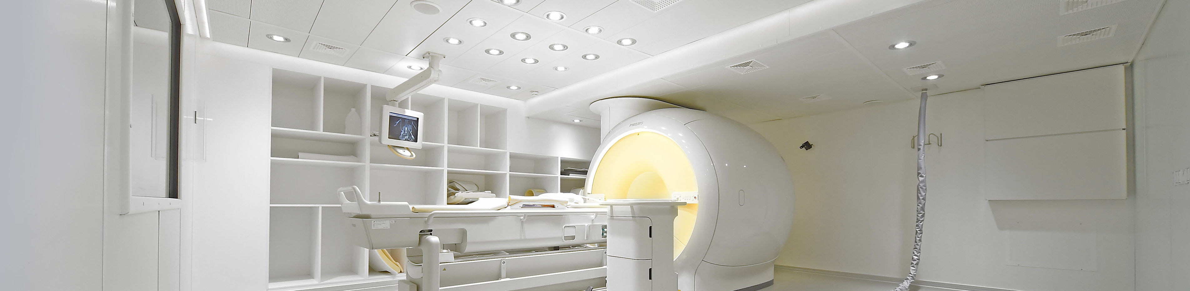
Cardiac MRI is a very safe examination technique. In contrast to computer tomography, MRI does not use X-rays to create images, but magnetic fields. For this reason, the duration of the examination is - depending on the problem - approx. 30 - 60 minutes longer than with computer tomography. During this time, you lie on the examination table in the MRI machine ("tube"). Cardiac MRIs are carried out by MTRs trained in cardiac MRI by the German Radiological Society at our institute in a state-of-the-art device (Philips Ingenia 1.5T) specially designed for cardiac imaging. This has a larger opening than older MRI machines, but claustrophobia ("claustrophobia") can rarely occur. If you suffer from claustrophobia, we can give you a sedative tablet before the start of the examination. Please note that in this case, active participation in road traffic is not permitted on the day of the examination. We do not have a so-called open MRI machine available due to the significantly poorer image quality in comparison. In order to obtain the sharpest possible images of the heart, it is important that you follow the breathing commands ("inhale, exhale, hold your breath") given by our assistants during the examination, as otherwise the heart would move up and down with the diaphragm and the images would not be usable, or only to a limited extent.
During the stress MRI, the heart is put into a "stress situation" for a short time (approx. 5 minutes) with the help of a drug (regadenoson). This is similar to the strain that the body feels during an endurance run. Accordingly, you will notice an acceleration of the pulse, a slight pressure on the chest and possibly a feeling of heat. These symptoms are completely normal and desirable, as they indicate the correct effect of the medication. You will be accompanied by a doctor during this "stressful situation". Immediately after administration of the regadenoson, the MR contrast medium is injected in order to detect a possible reduced blood flow to the heart muscle during the stress situation. To cancel out the effect of the regadenoson, we will give you dark chocolate with a high caffeine content, whereupon the symptoms described above usually subside within a few minutes of the administration of the medication.
After the end of the cardiac MRI, you can take part in everyday life as normal - even after a stress examination. The MRI examination is then evaluated and diagnosed by radiologists who are specially trained and experienced in cardiac imaging and who are certified in the field of cardiac MRI by the German Radiological Society.
If you would like to receive a CD or online access to the images from the examination, we will be happy to provide you with these after the examination. If you would like a final consultation with one of our radiologists to discuss the findings, this is of course possible. You will need to allow a little waiting time for this, as there are a large number of images to be evaluated (more than 1000), which must first be analyzed using special software. The final report is usually prepared on the same day and sent to your treating doctor, or to you personally if you wish.
Further information
- Information on our areas of focus
- Technical details about the equipment of our institute
- Find out more about magnetic resonance imaging research at our institute here