
Magnetic resonance imaging
Everything you need to know about the examination.
Magnetic resonance imaging is one of the most modern examination methods in diagnostic radiology, with which cross-sectional images of the human body can be produced in any desired plane without radiation exposure. Thanks to high resolution and high tissue contrast, anatomical structures can be visualized much better than with many other imaging techniques. In particular, different soft tissue structures can be better differentiated. To date, no damage has been reported due to the high magnetic flux density and electromagnetic high-frequency fields that occur during MRI.
If you have been referred to us for an MRI, you can find out about the examination procedure in advance here.
MRI-guided interventions
We also offer some procedures that do not fall into the area of MRI diagnostics/examinations, but use MRI technology to guide interventions. MRI scanners are primarily used to show what is happening in the body in real time and non-invasively. MRI technology therefore makes it possible to "look inside" the tissue without having to penetrate it. Find out more about:
Frequently asked questions
How can I prepare for the examination?
It is not necessary for you to be sober for a routine MRI examination. However, if this is necessary, you will be informed of this when you make your appointment.
If you are having an MRI examination with contrast medium, it is usually necessary for you to bring a current creatinine and TSH value with you, which you will have determined in advance (about a week before the examination) by your family doctor or the referring doctor with a blood test. These two values provide information about your kidney and thyroid function. You will be asked to provide these two values during your consultation with one of our radiologists. Without the information on these blood values, we are unfortunately unable to carry out the examination in your own interest, as this could otherwise result in damage to your health. If one of the two values is abnormal (i.e. not normal), please inform us in advance so that we can make the necessary decisions and plan the examination. If you have any questions, please contact us on the telephone number given above.
As the MRI works with strong magnetic fields, you are requested to remove all metal objects before entering the examination room, as this may otherwise result in injury. If you have metal implants in or on your body, please bring your implant passport with you - if you have one - and present it during the consultation. If you have a pacemaker, please inform us in advance, as a decision must first be made in cooperation with our cardiology colleagues as to whether and how an MRI examination is possible; special regulations apply here.
If you suffer from claustrophobia or are very overweight, please talk to us in advance. Depending on the degree of claustrophobia, it may also be possible to take a sedative tablet before the examination. However, if you come to the examination as an outpatient, you will then need to be collected. Due to the temporary restriction of your ability to react, you must not drive a motor vehicle or two-wheeler or work on dangerous machinery for 12 hours after the examination.
How does the examination work?
Before the examination, you will be informed by a radiologist about the benefits and risks of the examination. You will receive a detailed information sheet from us in preparation for the consultation. We ask you to read this carefully and complete it truthfully. You can then ask your questions during the subsequent consultation.
Some examinations require the intravenous administration of contrast medium. We only use the latest generation of contrast media, which are very well tolerated; only in rare cases can there be side effects, about which the doctor will inform you in detail. In order to administer the contrast medium, it is necessary for us to place a venous access in your arm (crook of the elbow or back of the hand) before the examination, which is similar to taking blood. The contrast medium is then administered via an automated pump during the examination. You may feel a little warm, which is completely normal.
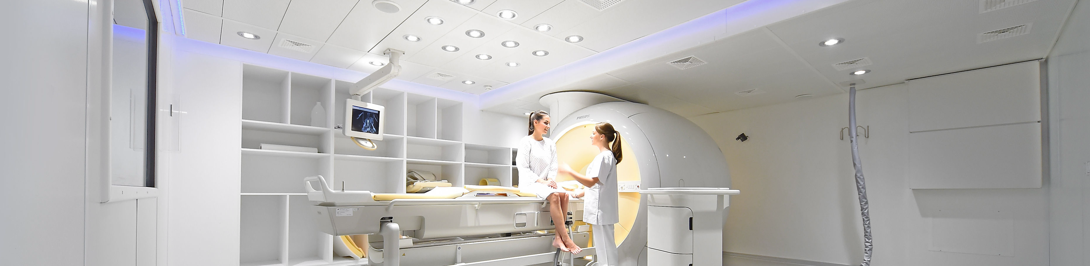
For the examination, the body region to be examined must be placed in the middle of a tube that is approx. 70 cm wide. The tube remains open during the entire examination, it is bright and well ventilated. The examination device is located in a shielded room and is guarded from the adjacent room by a window and a camera. You can speak to our staff via an intercom system during the examination. We can see you at all times during the examination so that nothing can happen to you.
The MRI examination takes about 20-45 minutes, depending on the problem. It takes this time for the measurements to be taken and for the computer to calculate the images. During this time, please lie as still as possible. You will hear loud, knocking noises during the examination; you will be given special headphones to protect your hearing. You will then be finished. If you would like to receive a CD of the examination, please wait a moment. If you would like to have a final consultation with one of our radiologists to discuss the findings, please also allow for a little waiting time, as the images must first be analyzed.
Retrieve images and findings digitally
After an examination at our institute, we will make your images available to you digitally via a QR code or access code. You can then view your images on a mobile device or on your computer. Your attending physician can also call up the images and discuss them with you.
In addition, the written report on your examination can be accessed via the online portal just a few hours after the examination. Of course, the written report will also be sent to the referring doctor by post.
To do this, scan the QR code you have received from us or enter the access code in the image portal of Cologne University Hospital.
For more detailed instructions, take a look at our flyer on the online web viewer.
Further information
- Information on our areas of focus
- Technical details about the equipment of our institute
- Find out more about magnetic resonance imaging research at our institute here