MRI arthrography
Direct arthrographies
At our institute, we offer MRI arthrography for the hip, shoulder and elbow. We usually carry out this special examination when a normal MRI was not sufficient to make an accurate diagnosis. These examinations are often performed to detect tears in the labrum (a cartilage structure).
Preparation:
- Before the examination, you should bring the following laboratory values from your GP: Thrombocytes, Quick, aPTT.
- Please inform us when you register if you are taking blood thinners.
- You do not need to be fasting, i.e. you can eat and drink normally beforehand.
Procedure of the examination:
First step - application of contrast medium: The examination begins with the injection of a contrast medium mixture into the joint. You will usually lie on your back. We use X-ray fluoroscopy to ensure that the contrast medium is placed correctly. Most patients do not find this painful. If necessary, you will be given a local anesthetic so that you do not feel any pain.
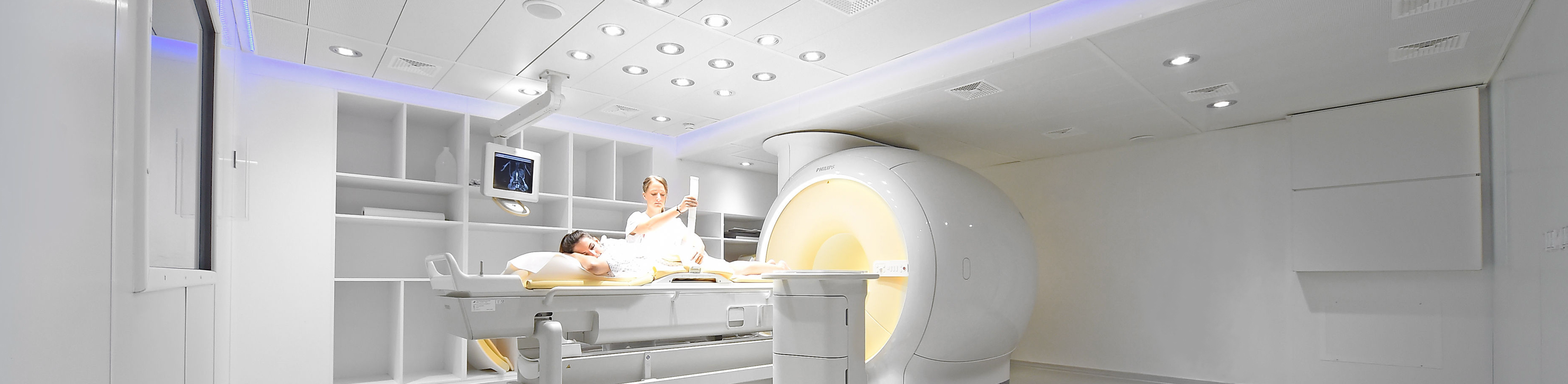
- Second step - carrying out the MRI: Once the contrast medium has been injected, the MRI examination follows. The contrast agent already applied helps us to better detect injuries and cracks that are not visible in a normal MRI.
Duration of the examination:
As the examination consists of two steps, it is somewhat more time-consuming. Please bring enough time with you, the entire examination usually takes about two hours.