
MR-Sellink
Everything you need to know about the examination
MR-Sellink is a special examination that uses magnetic resonance imaging (MRI ) and a contrast agent to visualize structures in the small intestine particularly well. This helps in the diagnosis of chronic inflammatory and tumorous diseases of the small intestine, such as Crohn's disease or colorectal cancer.
Frequently asked questions
How can I prepare for the examination?
The examination should be performed on an empty stomach, so do not eat anything on the day of the examination and 6 hours before the examination. Medication and drinks (without fat, strong acid or a lot of sugar - i.e. no fruit juices, soft drinks, milk or alcoholic drinks) can still be taken. If possible, you should stop eating high-fat and high-fiber foods such as whole grain products, legumes, salads, fruit and raw vegetables two days before the examination. On the day before the examination, please only eat a light breakfast and lunch and drink plenty of fluids in the afternoon and evening, if possible two liters of water and tea, no drinks with a lot of fat, strong acid or sugar - i.e. no fruit juices, soft drinks, milk or alcoholic drinks.
Immediately before the examination, you will be given a medication (Buscopan) to reduce intestinal activity so that the intestinal walls can be better assessed during the examination. If you suffer from increased intraocular pressure (glaucoma) or prostatic hyperplasia, please let us know during the consultation, as the administration of Buscopan is contraindicated in such cases. In addition, active participation in road traffic is no longer possible after the administration of Buscopan, as the medication can have an effect on vision for a certain period of time. For this reason, please make appropriate arrangements for your return journey in advance.
If you suffer from severe claustrophobia or are very overweight, please speak to us in advance. Depending on the severity of your claustrophobia, it may also be possible to take a sedative tablet before the examination. However, if you come to the examination as an outpatient, you will then need to be collected. Due to the temporary restriction of your ability to react, you must not drive a motor vehicle or two-wheeler or work on dangerous machinery for 12 hours after the examination.
How does the examination work?
For the MR-Sellink examination, you will be asked to drink two liters of an aqueous, special contrast medium solution (which contains cellulose) over a period of about one hour before the examination. This is distributed in the intestine during the examination and helps to better visualize the intestinal structures. Therefore, please arrive at the clinic at least 1.5 hours before the start of the examination.
Immediately before the start of the actual examination, you will be given a venous access through which the above-mentioned medication (Buscopan) will be administered.
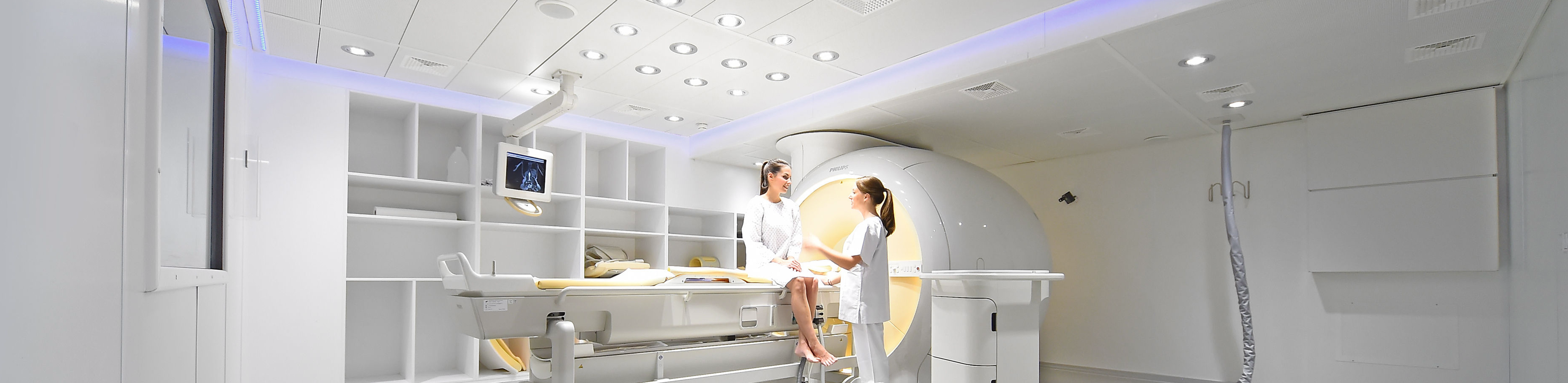
For the examination, the body region to be examined must be placed in the middle of a tube approximately 70 cm wide. The tube remains open during the entire examination, it is bright and well ventilated. The examination device is located in a shielded room and is guarded from the adjacent room by a window and a camera. You can speak to our staff via an intercom system during the examination. We can see you at all times during the examination so that nothing can happen to you.
The actual MRI examination takes about 30 minutes. It takes this time for the measurements to be taken and for the computer to calculate the images. During this time, please lie as still as possible. You will hear loud, knocking noises during the examination; you will be given special headphones to protect your hearing. You will then be finished. If you would like to receive a CD of the examination, please wait a moment. If you would like to have a final consultation with one of our radiologists to discuss the findings, please also allow for a little waiting time, as the images must first be analyzed.
Further information
- Information on our areas of focus
- Technical details about the equipment of our institute
- Find out more about magnetic resonance imaging research at our institute here