
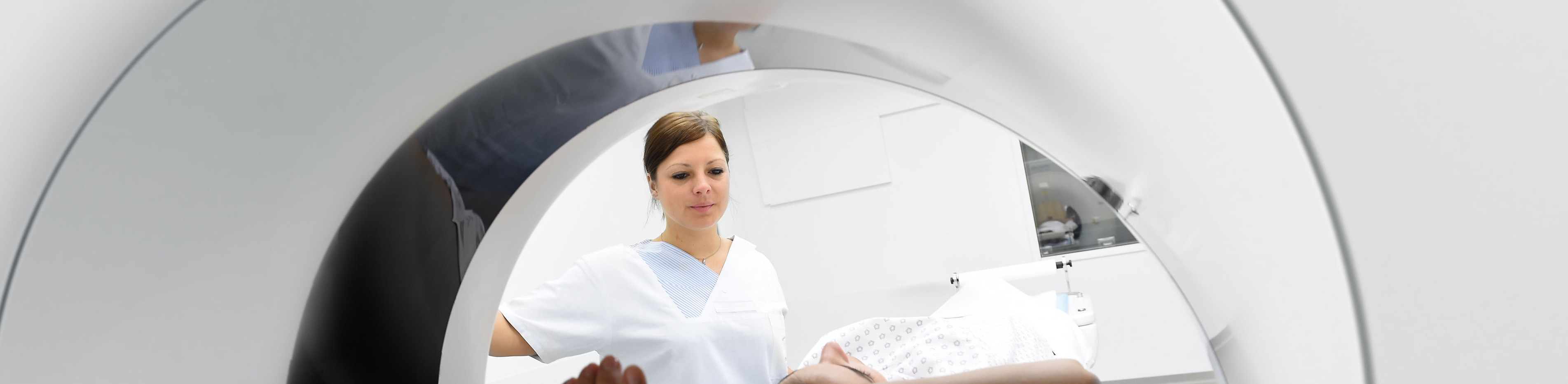
Cardiovascular imaging
Research topics
Cardiovascular computed tomography (CT)
A central aspect of our research in the field of cardiovascular computed tomography is the non-invasive characterization of pulmonary hypertension. In close collaboration with the Ludwig Boltzmann Institute in Graz, we are evaluating the added value of dual-layer dual-energy computed tomography (dlDECT) and knowledge-based algorithms for the diagnosis and prognosis of the disease. In addition, our group is dedicated to testing the technical and clinical potential of dlDECT imaging in a variety of cardiovascular diseases and conditions, such as acute pulmonary embolism, pre- and post-interventional imaging in TAVR patients and in-stent imaging of the coronary arteries. In addition, we are evaluating the use of deep learning models to improve the detection of cardiovascular diseases, e.g. intracranial aneurysms. We also aim to identify potential risk factors for cerebral ischemia using cardiac CT, focusing on left-sided septal pockets and diverticula.
Cardiovascular MRI
In the field of cardiovascular MRI, our research focus is on the evaluation of a novel 3D isotropic flow-independent MR angiography technique without contrast agent called Relaxation-enhanced angiography without contrast and triggering (REACT). We place particular emphasis on determining the efficacy of this technique in specific patient groups, including patients with Marfan syndrome, congenital heart defects and transplant recipients. In the area of cardiac MRI, we aim to expand clinical applicability through novel techniques to accelerate data acquisition, including compressed sensing, parallel imaging and artificial intelligence. This includes both functional (cine sequences) and morphologic techniques, in particular 3D isotropic late gadolinium enhancement imaging. In addition, we are investigating the use of large language models as a diagnostic tool in cardiac MRI.
Cooperations
- Clinic III for Internal Medicine, Heart Center, Faculty of Medicine and University Hospital Cologne, Cologne, Germany
- Clinic and Polyclinic for General, Visceral, Thoracic and Transplant Surgery, Heart Center, Faculty of Medicine and University Hospital Cologne, Cologne, Germany
- Ludwig Boltzmann Institute for Pulmonary Vascular Research, Graz, Austria
- Clinic for Pneumology, University of Graz, Graz, Austria
- Clinic for Diagnostic and Interventional Radiology, University Hospital Bonn, Bonn, Germany
- Department of Radiology, Diagnostic and Interventional Radiology, University of Tübingen, Tübingen, Germany
- Clinic for Diagnostic and Interventional Radiology, University Hospital of Johannes Gutenberg University, Mainz, Germany
- Department of Radiology, Neuroradiology and Nuclear Medicine, Johannes Wesling University Hospital, Ruhr University Bochum, Germany
Promotion
- Funded by the Cologne Clinician Scientist Program (CCSP) / Faculty of Medicine / University of Cologne. Funded by the German Research Foundation (DFG) (project no. 413543196)
- The position as Clinician Scientist is supported by the Dean's Office of the Faculty of Medicine and the University Hospital Cologne.
Selected publications
Advanced CT measures of coronary artery disease with intermediate stenosis in patients with severe aortic valve stenosis. Eur Radiol. 2024
Dual-layer dual-energy CT-derived pulmonary perfusion for the differentiation of acute pulmonary embolism and chronic thromboembolic pulmonary hypertension. Eur Radiol. 2023
Reduction of contrast medium for transcatheter aortic valve replacement planning using a spectral detector CT: a prospective clinical trial. Eur Radiol. 2023
Coronary calcium scoring using virtual non-contrast reconstructions on a dual-layer spectral CT system: Feasibility in the clinical practice. Eur J Radiol. 2023
Association between pouch morphology, cardiovascular risk factors and ischemic brain lesions in patients with left-sided septal pouches. Clin Imaging. 2023
Imaging diagnostics in pulmonary hypertension. Radiology up2date. 2023
German Radiologists Position Paper on Coronary computed tomography: Clinical Evidence and Quality of Patient Care in Chronic Coronary Syndrome. Rofo. 2023
Preexisting Chronic Thromboembolic Pulmonary Hypertension in Acute Pulmonary Embolism. Chest. 2023
Spectral Detector CT-Derived Pulmonary Perfusion Maps and Pulmonary Parenchyma Characteristics for the Semiautomated Classification of Pulmonary Hypertension. Front Cardiovasc Med. 2022
The Aortic Ductus Diverticulum-Innocent Bystander or Potential Source of Thromboembolic Stroke? J Comput Assist Tomogr. 2022
Detection of patients with chronic thromboembolic pulmonary hypertension by volumetric iodine quantification in the lung-a case control study. Quant Imaging Med Surg. 2022
Imaging of the Left Atrial Appendage Before Occluder Device Placement: Evaluation of Virtual Monoenergetic Images in a Single-Bolus Dual-Phase Protocol. J Comput Assist Tomogr. 2022
Feasibility and Comparison of Resting Full-Cycle Ratio and Computed Tomography Fractional Flow Reserve in Patients with Severe Aortic Valve Stenosis. J Cardiovasc Dev Dis. 2022
Reduction of CT artifacts from cardiac implantable electronic devices using a combination of virtual monoenergetic images and post-processing algorithms. Eur Radiol. 2021
Deep learning assistance increases the detection sensitivity of radiologists for secondary intracranial aneurysms in subarachnoid haemorrhage. Neuroradiology. 2021
Abdominal vessel depiction on virtual triphasic spectral detector CT: initial clinical experience. Abdom Radiol (NY). 2021
Fully Automated Detection and Segmentation of Intracranial Aneurysms in Subarachnoid Hemorrhage on CTA Using Deep Learning. Sci Rep 2020
Are left atrial diverticula and left-sided septal pouches relevant additional findings in cardiac CT? Correlation between left atrial outpouching structures and ischemic brain alterations. Int J Cardiol. 2020
Virtual monoenergetic images and post-processing algorithms effectively reduce CT artifacts from intracranial aneurysm treatment. Sci Rep. 2020
Photon-Counting Computed Tomography for Coronary Stent Imaging: Evaluation of the Potential Clinical Impact for the Delineation of In-Stent Restenosis. Invest Radiol. 2020
Evaluation of soft-plaque stenoses in coronary artery stents using conventional and monoenergetic images: first in-vitro experience and comparison of two different dual-energy techniques. Quant Imaging Med Surg. 2020
Knowledge-based iterative reconstructions for imaging of coronary artery stents: first in-vitro experience and comparison of different radiation dose levels and kernel settings. Acta Radiol. 2019
Software-automated multidetector computed tomography-based prosthesis-sizing in transcatheter aortic valve replacement: Inter-vendor comparison and relation to patient outcome. Int J Cardiol. 2018
Fourth update on CT angiography of coronary stents: in vitro evaluation of 24 novel stent types Acta Radiol. 2018
Influence of spectral detector CT based monoenergetic images on the computer-aided detection of pulmonary artery embolism. Eur J Radiol. 2017
Monoenergetic reconstructions for imaging of coronary artery stents using spectral detector CT: In-vitro experience and comparison to conventional images. J Cardiovasc Comput Tomogr. 2017
Non-invasive imaging of bioresorbable coronary scaffolds using CT and MRI: First in vitro experience. Int J Cardiol. 2016
Coronary artery stent imaging with CT using an integrated electronics detector and iterative reconstructions: first in vitro experience. J Cardiovasc Comput Tomogr. 2013
Multiparametric Monitoring of Disease Progression in Contemporary Patients with Wild-Type Transthyretin Amyloid Cardiomyopathy Initiating Tafamidis Treatment. J Clin Med 2024
Compressed SENSE accelerated 3D single-breath-hold late gadolinium enhancement cardiovascular magnetic resonance with isotropic resolution: clinical evaluation. Front Cardiovasc Med. 2023
Thoracic aorta diameters in Marfan patients: Intraindividual comparison of 3D modified relaxation-enhanced angiography without contrast and triggering (REACT) with transthoracic echocardiography. Int J Cardiol. 2023
GPT-4 for Automated Determination of Radiologic Study and Protocol Based on Radiology Request Forms: A Feasibility Study. Radiology. 2023
Imaging modalities in early cardiac transthyretin amyloidosis: who is first? Clin Res Cardiol. 2023
Structured Reporting in Cross-Sectional Imaging of the Heart: Reporting Templates for CMR Imaging of Ischemia and Myocardial Viability and for Cardiac CT Imaging of Coronary Heart Disease and TAVI Planning. Rofo. 2023
Imaging of the extracranial internal carotid artery in acute ischemic stroke: assessment of stenosis, plaques, and image quality using relaxation-enhanced angiography without contrast and triggering (REACT). Quant Imaging Med Surg. 2022
Medium-Term Outcomes of the HFpEF Stress Trial. JACC Cardiovasc Imaging. 2022
Defining the optimal temporal and spatial resolution for cardiovascular magnetic resonance imaging feature tracking. J Cardiovasc Magn Reson. 2021
k-t accelerated multi-VENC 4D flow MRI improves vortex assessment in pulmonary hypertension. Eur J Radiol. 2021
Velocity quantification in 44 healthy volunteers using accelerated multi-VENC 4D flow CMR. Eur J Radiol. 2021
Exercise Stress Real-Time Cardiac Magnetic Resonance Imaging for Noninvasive Characterization of Heart Failure With Preserved Ejection Fraction: The HFpEF-Stress Trial. Circulation. 2021
Relaxation-Enhanced Angiography Without Contrast and Triggering (REACT) for Fast Imaging of Extracranial Arteries in Acute Ischemic Stroke at 3 T. Clin Neuroradiol. 2021
Technique and clinical significance of cardiac mapping - what the radiologist should know. Radiology up2date. 2021
Comparison of a novel Compressed SENSE accelerated 3D modified relaxation-enhanced angiography without contrast and triggering with CE-MRA in imaging of the thoracic aorta. Int J Cardiovasc Imaging. 2021
Clinical application of free-breathing 3D whole heart late gadolinium enhancement cardiovascular magnetic resonance with high isotropic spatial resolution using Compressed SENSE. J Cardiovasc Magn Reson. 2020
Imaging of the pulmonary vasculature in congenital heart disease without gadolinium contrast: Intraindividual comparison of a novel Compressed SENSE accelerated 3D modified REACT with 4D contrast-enhanced magnetic resonance angiography. J Cardiovasc Magn Reson. 2020
Multiparametric MRI of the heart - common and rare findings. Rofo. 2020
Structured Reporting in Cross-Sectional Imaging of the Heart: Reporting Templates for CMR Imaging of Cardiomyopathies (Myocarditis, Dilated Cardiomyopathy, Hypertrophic Cardiomyopathy, Arrhythmogenic Right Ventricular Cardiomyopathy and Siderosis). Rofo. 2020
Atrial mechanics and their prognostic impact in Takotsubo syndrome: a cardiovascular magnetic resonance imaging study. Eur Heart J Cardiovasc Imaging. 2019
Precision, reproducibility and applicability of an undersampled multi-venc 4D flow MRI sequence for the assessment of cardiac hemodynamics. Magn Reson Imaging. 2019
Cardiovascular magnetic resonance imaging feature tracking: Impact of training on observer performance and reproducibility. PLoS One. 2019
Large Saccular Aneurysm at the Ostium of the Coronary Sinus Mimicking a Right-Atrial Myxoma. Circ Cardiovasc Imaging. 2018
Inter-vendor reproducibility of left and right ventricular cardiovascular magnetic resonance myocardial feature-tracking. PLoS One. 2018
Myocardial T1 and T2 mapping in severe aortic stenosis: Potential novel insights into the pathophysiology of myocardial remodeling. Eur J Radiol. 2018
Incremental value of cardiovascular magnetic resonance feature tracking derived atrial and ventricular strain parameters in a comprehensive approach for the diagnosis of acute myocarditis. Eur J Radiol. 2018
In vitro evaluation of flow patterns and turbulent kinetic energy in trans-catheter aortic valve prostheses. MAGMA. 2018
A novel multiparametric imaging approach to acute myocarditis using T2-mapping and CMR feature tracking. J Cardiovasc Magn Reson. 2017
Re-evaluation of a novel approach for quantitative myocardial oedema detection by analyzing tissue inhomogeneity in acute myocarditis using T2-mapping. Eur Radiol. 2017
Intra- and inter-observer reproducibility of global and regional magnetic resonance feature tracking derived strain parameters of the left and right ventricle. Eur J Radiol. 2017
Left and right atrial feature tracking in acute myocarditis: A feasibility study. Eur J Radiol. 2017
Modern Imaging of Myocarditis: Possibilities and Challenges. Rofo. 2016
Reproducibility of three different cardiac T2-mapping sequences at 1.5T. J Magn Reson Imaging. 2016
Diagnostic implications of magnetic resonance feature tracking derived myocardial strain parameters in acute myocarditis. Eur J Radiol. 2016
Mapping tissue inhomogeneity in acute myocarditis: a novel analytical approach to quantitative myocardial edema imaging by T2-mapping. J Cardiovasc Magn Reson. 2015
Quantitative Analysis of Vortical Blood Flow in the Thoracic Aorta Using 4D Phase Contrast MRI. PLoS One. 2015
A systematic evaluation of three different cardiac T2-mapping sequences at 1.5 and 3T in healthy volunteers. Eur J Radiol. 2015
Cardiac T2-mapping using a fast gradient echo spin echo sequence - first in vitro and in vivo experience. J Cardiovasc Magn Reson. 2015
Biventricular myocardial strain analysis in patients with arrhythmogenic right ventricular cardiomyopathy (ARVC) using cardiovascular magnetic resonance feature tracking. J Cardiovasc Magn Reson. 2014
4D phase contrast flow imaging for in-stent flow visualization and assessment of stent patency in peripheral vascular stents--a phantom study. Eur J Radiol. 2012
Feasibility of functional cardiac MR imaging in mice using a clinical 3 Tesla whole body scanner. Invest Radiol. 2009
Team
Dr. Lukas Goertz, MD
Prof. Dr. Dr. Nils Große Hokamp, MD
Dr. Jan Paul Janßen, MD
Mr. Kenan Kaya, MD
Dr. Konstantin Klein, MD
Dr. Jonathan Kottlors, MD
Dr. Isabel Langenbach, MD
Priv.-Doz. Dr. Simon Lennartz, MD
Vanessa Schmidt
Dr. Thomas Schömig, MD
Univ.-Prof. Dr. Stephan Rosenkranz, Clinic III for Internal Medicine, Heart Center, Faculty of Medicine and University Hospital Cologne, Cologne, Germany
Prof. Dr. Roman Pfister, Clinic III for Internal Medicine, Heart Center, Faculty of Medicine and University Hospital Cologne, Cologne, Germany
Priv.-Doz. Dr. Henrik ten Freyhaus, Clinic III for Internal Medicine, Heart Center, Faculty of Medicine and University Hospital Cologne, Cologne, Germany
Dr. Katharina Seuthe, Clinic III for Internal Medicine, Heart Center, Faculty of Medicine and University Hospital Cologne, Cologne, Germany
Priv.-Doz. Dr. Christopher Hohmann, Clinic III for Internal Medicine, Heart Center, Faculty of Medicine and University Hospital Cologne, Cologne, Germany
Univ.-Prof. Dr. Horst Olschievski, Deputy Director of the Ludwig Boltzmann Institute for Pulmonary Vascular Research, Head of the Department of Pneumology, University of Graz, Graz, Austria
Dr. Michael Pienn, PhD, Dipl. Ing., Ludwig Boltzmann Institute for Pulmonary Vascular Research, Graz, Austria
Prof. Dr. Claas Philip Nähle, Radiological Alliance, Hamburg, Germany
Priv.-Doz. Dr. Erkan Celik, Institute for Diagnostic and Interventional Radiology, Medical Faculty and University Hospital Cologne, Cologne, Germany
Dr. Marcel C. Langenbach, Institute of Diagnostic and Interventional Radiology, Faculty of Medicine and University Hospital Cologne, Cologne, Germany
Prof. Dr. Jan Borggrefe, Johannes Wesling University Hospital, Ruhr University Bochum, Department of Radiology, Neuroradiology and Nuclear Medicine, Germany
Dr. Jan-Robert Kröger, Johannes Wesling University Hospital, Ruhr University Bochum, Department of Radiology, Neuroradiology and Nuclear Medicine, Germany
Dr. Florian Fintelmann, Massachusetts General Hospital, Department of Radiology, Boston, USA, Head of Thoracic Imaging, Percutaneous Thermal Ablation, Associate Professor
Prof. Dr. Christoph Kabbasch, Institute for Diagnostic and Interventional Radiology, Faculty of Medicine and University Hospital Cologne, Cologne, Germany
Dr. Carsten Herbert Gietzen, Institute of Diagnostic and Interventional Radiology, Faculty of Medicine and University Hospital Cologne, Cologne, Germany
Dr. Kilian Weiss, PhD, Philips GmbH
Mr. Christoph Katemann, Philips GmbH
Dr. Kristina Sonnabend, PhD, Philips GmbH
Further information
- Information on our areas of focus
- Technical details about the equipment at our institute
- Find out more about cardiovascular imaging in clinical practice at our institute here