
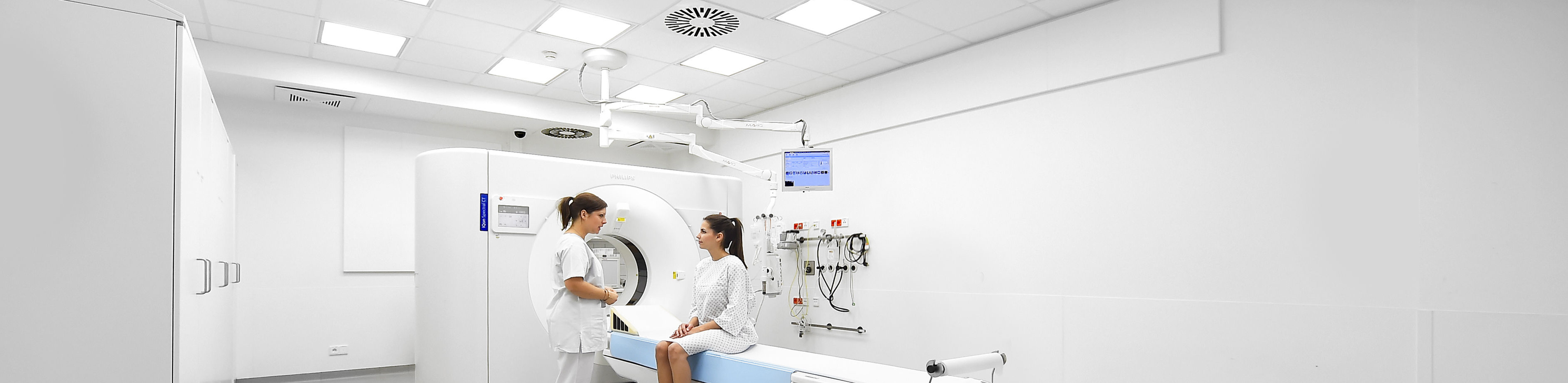
Spectral detector CT
Spectral-detector computed tomography, or spectral CT for short, is a new type of technology that supplements conventional CT examinations with spectral image data. It is the latest development of dual-energy CT (DECT for short). The independent registration of high- and low-energy X-rays enables improved image quality and more detailed tissue characterization - without increasing radiation exposure!
As the first radiology facility in Europe and one of the first clinics in the world to use spectral CT clinically, we have had extensive experience with this technology since it was first installed in 2016. With the commissioning of a second-generation spectral CT scanner in September 2022, its use was further expanded and has since been used almost across the board in routine clinical care.
Spectral CT enables significantly improved image quality with reduced radiation exposure and optimized contrast agent administration. It also allows new types of image reconstruction for the differentiated assessment of unclear incidental findings and for precise material characterization, for example in the case of kidney stones or in gout diagnostics.
The Institute of Diagnostic and Interventional Radiology at Cologne University Hospital is an internationally recognized reference center for spectral CT. Our experience and research results are regularly presented at national and international congresses(computed tomography research).
Further information
- Information on our areas of focus
- Technical details about the equipment of our institute
- Find out more about computed tomography research at our institute here